")
")

An X-ray microscope is an instrument used to produce enlarged images of samples illuminated with X-rays. There are two main principles of microscopes to be distinguished: full field microscopes and scanning microscopes. In full field microscopes the whole field of view is imaged to a detector plane at the same time. In scanning microscopes the sample is illuminated with a bright well focused spot scanning over the sample. The detector then measures the total intensity over time coming from the currently illuminated spot on the sample and the image is calculated from this data when the scanning process is finished.
What are the advantages of X-ray microscopes over other types of microscopes?
X-rays penetrate matter far easier than visible light. So X-ray microscopes can image the inside of samples opaque for visible light.
X-ray microscopes can achieve higher optical resolution than microscopes using visible light. The wavelength of X-rays is much shorter than the wavelength of visible light, so the limit of the optical resolution (caused by diffraction) of X-ray microscopes is far below the diffraction limit of microscopes working with visible light. Scanning electron microscopes in comparison have a high image resolution, but they need vacuum-proof samples with metallic or metallized surfaces and they can not image the inner of a sample. In table 1 some characteristics of different microscopes are compared.
| X-ray microscope | Visible light microscope | Scanning electron microscope | |
| Wavelength range | ≈0.03 nm to 10 nm | 400 nm to 800 nm | ≈0.06 nm to 3 nm |
| Optical resolution reached | ≈20 nm | ≈200 nm | ≈2 nm |
| Imaging | inner of sample | inner/surface of sample | surface of sample |
| Depth of field (relative [Hil 1956]) |
medium to high (25) |
low (1) | high (570) |
| Possible samples | all | all | vacuum proof; metallic or metallized |
| Imaging modes | scanning/full field | (scanning)/full field | scanning |
Table 1: Comparing different types of microscopes (values only for orientation)
Full field X-ray microscopes can be either enlarging shadow projection microscopes (figure 1) or microscopes with focusing optical elements (figure 2).
Shadow projection microscopes
consist of a point-shaped X-ray source, optional of an optics 4 producing a demagnified image 5 of the source 1, the sample 2 positioned close to the source 1 (respectively to the image 5 of the source) and a detector for the resulting X-ray image 3 (fig. 1). Influencing the direction of the X-rays is not needed in the basic version of this type of microscope.
The magnification of the set-up is the quotient L2 / L1 with the distance source-sample L1 and the distance source-image L2. The resulting image resolution mainly depends on the diameter DSource of the X-ray source, as the half shades caused by a larger source lead to blurred images. Two points on the sample in a minimum distance of about DSource can be resolved with this type of microscope. This limitation can be reduced using a focusing optics 4 in front of the sample. It creates a demagnified image of the source. This image of the source is used as source for the shadow projection microscope. Physically the maximum image resolution is limited by diffraction.
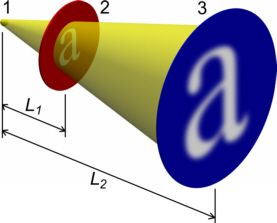
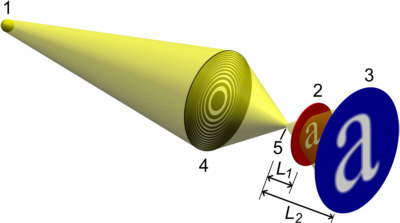
Fig. 1: Shadow projection microscope set-up producing enlarged images of a sample: with a small source 1 (left) and using a reduced size image 5 (using e.g. a Fresnel zone plate) of the source 1 for sharper images (right)
Microscopes with focusing optical elements
consist of an X-ray source, optional of an illumination optics, the sample, an imaging lens or optics, a detector for the resulting X-ray image and an optical bench as a basis to align the components.
The source can be an X-ray tube, a synchrotron or a plasma source. The intensity of the source determines the time needed to take an image with a good signal to noise ratio. Depending on the type of microscope the spectral distribution of the photon energy of the X-rays has to fit the optical properties of the microscope. Mostly a small photon energy range is required for proper function of the overall system.
The purpose of the illumination optics is to collect as many photons as possible and to deliver them on the sample. This helps reducing the acquisition time for an image. Mostly a rectangular area on the sample should be homogeneously illuminated. The illumination optics has to be capable to concentrate the incoming X-ray beam in respect to its spectral photon energy distribution. But also it has to fullfil requirements concerning the angular distribution of the beam delivered at the sample, to guarantee an optimal overall imaging quality of the microscope.
The sample is fixed in a sample holder. This holder (or the stage it is mounted on) mostly is motorized to allow for precise positioning of the sample in the X-ray beam, to choose a section of interest and to be able to focus and sometimes to adjust the magnification of the system.
The imaging lens is positioned in a distance of more than its focal length (and less than twice its focal length for magnified imaging) from the sample. Its purpose is to image each point of the sample (within the field of view) to a corresponding point in the image plane. The imaging lens has to be suited for the photon energy used and its entrance acceptance angle needs to be large enough to image the desired field of view.
The function of the image X-ray detector is to convert the X-ray intensity distribution in the image plane into a visible image or into electrical signals that are displayed as an image on a computer monitor. So the detector can be a photographic film, an image plate detector or a scintillator crystal converting the X-rays into visible light in combination with a CCD-detector capturing the visible light image.
An inflexible optical bench to mount the components of the microscope on top is a necessary prerequisite when a high optical resolution is aimed for.

Fig. 2: Sketch of an example set-up of a full field hard X-ray microscope, in this example using only refractive X-ray lenses ©01
Scanning X-ray microscopes consist of an X-ray source, an optics to produce a high intensity spot on the sample, a scanning stage holding the sample, a detector measuring the incoming photon flux and an optical bench as a basis to align the components.
Source and optical bench are analogue to the components in a full field microscope.
The focal spot generating optics can e.g. be a capillary optics, a Fresnel zone plate, a compound refractive lens or a mirror optics. The main demand to this optics is to bring a maximum of photons into a minimum spot size on the sample. If the sample is rather thick, the optics should generate a focused beam with a long waist along the optical axis. If the waist of the double cone shaped beam is very short, the resulting image quality will be reduced.
The scanning stage moves the sample through the beam, line by line until the whole field of view is covered. The required step size of the stage depends on the desired image resolution. As a guideline, the step size should be about half the focal spot size to reach maximum possible image quality. The position of the stage has to be controlled better than the step size and the time dependend stage position has to be registered in order to render the image reconstruction possible.

Fig. 3: Sketch of an example set-up of a scanning X-ray microscope, in this example using a Fresnel zone plate (with its necessary order sorting apertures)
| [Hil 1956] | Günter Hildenbrand, Röntgenoptik und Röntgenmikroskopie, Fortschritte der Physik, vol. 4, pp. 1-32, 1956 |